16-OH-E1, or 16-hydroxyestrone, is one of three routes your body can take when it breaks estrone down in the first phase of estrogen detoxification. It is the most estrogenic of the three, binding to estrogen receptors more strongly than 2-OH-E1 or 4-OH-E1, which means it can drive symptoms of high estrogen even when your overall estrogen sits in range.
Your result is read as a percentage of all three Phase I metabolites as well as a number on its own. If yours came back high, that is common and it responds well to the right support, and your practitioner can shift more of your estrogen onto the safer 2-OH-E1 pathway.
If you have not tested yet, 16-OH-E1 is measured in all three FUTURE WOMAN tests.
Let’s take a closer look at what 16-OH-E1 is, why optimal levels matter, and how they can impact your health.
What is 16-OH-E1?
16-OH-E1, or 16-hydroxyestrone, is a metabolite of estrogen. Estrogen is converted into three primary forms: estrone (E1), estradiol (E2), and estriol (E3), through the action of CYP enzymes. During Phase 1 estrogen detoxification, estrone (E1) is metabolised into 2-hydroxyestrone (2-OH-E1), 4-hydroxyestrone (4-OH-E1), or 16-hydroxyestrone (16-OH-E1) via distinct biochemical pathways. Essentially 2-OH-E1, 4-OH-E1 and 16-OH-E1 show us how estrogen moves out of the body.
- 2-OH-E1: Generally recognised as the safer metabolite. It weakly stimulates cell proliferation and may even inhibit it.
- 4-OH-E1: This metabolite can damage DNA, making it potentially more carcinogenic.
- 16-OH-E1: Binds to estrogen receptors with greater strength than other metabolites, making it more proliferative, pro-inflammatory, and angiogenic (encouraging new blood vessel growth).

Among these, the 16-OH-E1 pathway is considered the most estrogenic, meaning it may lead to estrogenic symptoms even when levels of general estrogens are not particularly elevated.
Estrogens exert their effects by binding to estrogen receptors, found in various tissues, including:
- Reproductive tissues: e.g., the endometrium.
- Non-reproductive tissues: e.g., bone, breast tissue, and the brain.
When 16-OH-E1 binds to these receptors, its strong activity can lead to increased cell division and inflammation, highlighting its role in certain health conditions.
Which pathway your body favours matters more when there is more estrogen to clear, so if you are in perimenopause, when estrogen fluctuates up and down, our guide to the perimenopause hormone test covers what to test and when.
Why Are High Levels of 16-OH-E1 Significant?
Elevated levels of 16-OH-E1 have been linked to several health concerns, including:
- Increased hormonal symptoms: High 16-OH-E1 is linked to increased symptoms such as heavy periods, painful periods and PMS.
- Hormone-dependent cancers: High 16-OH-E1 levels are potentially associated with breast and endometrial cancers.
- Endometrial conditions: The pro-inflammatory and proliferative nature of high 16-OH-E1 levels may be associated with endometriosis and fibroid growth.
- Cardiovascular disease (CVD): In postmenopausal women, increased levels of 16-OH-E1 are linked to a higher risk of CVD. Research suggests its potent estrogenic activity may influence vascular and endothelial function, contributing to cardiovascular risks.
- Autoimmune conditions: Elevated urinary 16-OH-E1 metabolites are often seen in patients with rheumatoid arthritis (RA) or systemic lupus erythematosus.
- Idiopathic Pulmonary Arterial Hypertension (iPAH):
In iPAH, 16-OH-E1 metabolites are thought to increase inflammatory cytokines, stimulate blood vessel growth (angiogenesis) and promote cellular proliferation.
High 16-OH-E1 is not all downside. Its estrogenic activity appears to offer some protection against osteoporosis, which is part of why this marker is read alongside the others rather than judged on its own.
16-OH-E1 and Breast Cancer Risk
Research has demonstrated that 16-OH-E1 plays a role in breast cancer development. Findings showed that levels of 16-OH-E1 were eight times higher in cancerous breast tissue compared to nearby healthy tissue. This suggests that 16-OH-E1 production may contribute to breast cancer induction. Luckily we can support moving the preference from 16-OH-E1 to 2-OH-E1 through your personalised plan.
Symptoms of High and Low 16-OH-E1 Levels
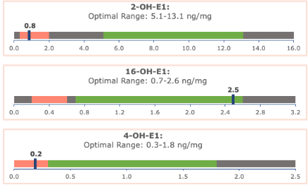
The luteal phase range for 16-OH-E1 is 0.7 – 2.6 ng/mg and the post menopausal range is 0.2-0.6 ng/mg.
16-OH-E1 are also reported as a percentage of all 3 Phase I metabolites. The expected and preferred percentages in women are;
- 2-OH-E1 60-80%
- 4-OH-E1 7.5-11%
- 16-OH-E1 13-30%
High 16-OH-E1 Levels:
Elevated 16-OH-E1 can lead to symptoms of estrogen dominance, due to its estrogenic effect, including:
- Breast tenderness
- Bloating
- Mood swings
- Heavy periods
- Weight gain
In women undergoing Hormone Replacement Therapy (HRT), favouring the 16-OH-E1 pathway can worsen symptoms such as:
- Breast pain
- Heavy bleeding
- Night sweats
- Hot flushes
Low 16-OH-E1 Levels:
While low levels of 16-OH-E1 are less concerning, they can result in symptoms of low estrogen, such as:
- Vaginal dryness
- Hot flashes
- Decreased libido
Low levels may also negatively affect bone health. A comprehensive interpretation of 16-OH-E1 levels requires evaluating them alongside other estrogen metabolites and detoxification pathways.
Which tests measure 16-OH-E1?
16-OH-E1 is measured in all three of our tests: the Classic Hormone Test, the Advanced Hormone Test and the Advanced+ Hormone Test with Cycle Mapping.
If you are not sure which test you need, our comparison guide walks through the differences.
Next steps
If your results suggest high or low 16-OH-E1 levels, this will be addressed in your personalised health plan by one of our experienced nutritionists. If you would like to learn more about your test results, remember you can purchase a 45 minute Hormone Health Consultation with your practitioner to discuss your results in more detail.
References
Correa, L. F., Zheng, Y., Delaney, A. A., Khan, Z., Shenoy, C. C., & Daftary, G. S. (2016). TGF-β Induces Endometriotic Progression via a Noncanonical, KLF11-Mediated Mechanism. Endocrinology, 157(9), 3332–3343. https://doi.org/10.1210/en.2016-1194
Denver, N., Homer, N. Z. M., Andrew, R., Harvey, K. Y., Morrell, N., Austin, E. D., & MacLean, M. R. (2020). Estrogen metabolites in a small cohort of patients with idiopathic pulmonary arterial hypertension. Pulmonary circulation, 10(1), 2045894020908783. https://doi.org/10.1177/2045894020908783
Khan W. A. (2019). 16 α-Hydroxyestrone induced adduct generate high affinity autoantibodies in SLE. Advances in medical sciences, 64(1), 72–78. https://doi.org/10.1016/j.advms.2018.11.002
Yu, K., Huang, Z. Y., Xu, X. L., Li, J., Fu, X. W., & Deng, S. L. (2022). Estrogen Receptor Function: Impact on the Human Endometrium. Frontiers in endocrinology, 13, 827724. https://doi.org/10.3389/fendo.2022.827724
Zheng, Y., Tabbaa, Z. M., Khan, Z., Schoolmeester, J. K., El-Nashar, S., Famuyide, A., Keeney, G. L., & Daftary, G. S. (2014). Epigenetic regulation of uterine biology by transcription factor KLF11 via posttranslational histone deacetylation of cytochrome p450 metabolic enzymes. Endocrinology, 155(11), 4507–4520. https://doi.org/10.1210/en.2014-1139
Zhu, B. T., Han, G. Z., Shim, J. Y., Wen, Y., & Jiang, X. R. (2006). Quantitative structure-activity relationship of various endogenous estrogen metabolites for human estrogen receptor alpha and beta subtypes: Insights into the structural determinants favoring a differential subtype binding. Endocrinology, 147(9), 4132–4150. https://doi.org/10.1210/en.2006-0113Ziegler, R. G., Fuhrman, B. J., Moore, S. C., & Matthews, C. E. (2015). Epidemiologic studies of estrogen metabolism and breast cancer. Steroids, 99(Pt A), 67–75. https://doi.org/10.1016/j.steroids.2015.02.015